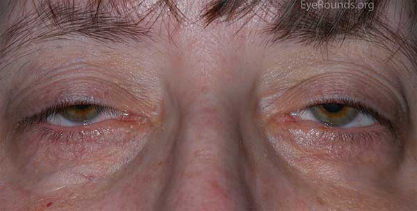
Myotonic Dystrophy Droopy Eyelid Symptoms
Many of the individuals with myotonic dystrophy run into issues with Drooping Eyelids. Here is some information from Eyeptosis.com
Patients present with bilateral ptosis and a flat facial expression. Myotonic dystrophy can be distinguished from oculopharyngeal dystrophy by the associated systemic findings such as “Christmas tree” cataracts, frontal balding, intellectual impairment, and heart block. In addition, the orbicularis weakness and ocular motility deficits are more prominent in myotonic dystrophy compared to OPMD. Diagnosis is confirmed by genetic testing for an expanded CTG repeat in the DMPK gene.
The disorder known as ptosis of the eye can be caused by many contributing factors including, the condition of myotonic dystrophy, an allergy, botulism, a brain tumor, the onset of diabetes, bell’s palsy or other nerve problems that affect the face, Guillain-Barre syndrome, chronic migraine, Multiple Sclerosis (MS), myasthenia gravis, a stroke, a stye, an abscess, an infection caused by bacteria, Cobra venom, eye or orbital tumor, and Horner syndrome. Other causes might also include other sources of nerve damage, aging, and a change in anatomy, recent eye surgery, nerve palsy, or trauma.
Muscular dystrophy is one of the many causes of Ptosis; therefore, both of these conditions are directly related. Myotonic Dystrophy is a continual wasting of the muscles and because Ptosis of the eye refers to weakening of the muscles as a direct result of the nerve endings not properly working, the muscular dystrophy has the ability to cause this drooping eyelid condition. The muscular dystrophy spreads throughout the body, weakening muscles in its path. This condition can commonly affect several of the body’s muscles from working properly, which is primarily what the condition Ptosis is characterized by (weakening of the eye and eyelid muscles, preventing the proper function) Therefore, muscular dystrophy can indirectly cause the droopy eye condition known as Ptosis.
For more information go to Eyeptosis.com
How is droopy eyelid treated?

The treatment for droopy eyelid depends on the specific cause and the severity of the ptosis.
If the condition is the result of age or something you were born with, your doctor may explain that nothing needs to be done because the condition isn’t usually harmful to your health. However, you may opt for plastic surgery if you want to reduce the drooping.
If your doctor finds that your droopy eyelid is caused by an underlying condition, you will likely be treated for that. This should typically stop the eyelids from sagging.
If your eyelid blocks your vision, you’ll need medical treatment. Your doctor may recommend surgery.
Glasses that can hold the eyelid up, called a ptosis crutch, are another option. This treatment is often most effective when the droopy eyelid is only temporary. Glasses may also be recommended if you aren’t a good candidate for surgery.
Surgery
Your doctor may recommend ptosis surgery. During this procedure, the levator muscle is tightened. This will lift the eyelid up into the desired position. For children who have ptosis, doctors sometimes recommend surgery to prevent the onset of lazy eye (amblyopia).
However, there are risks associated with surgery, including dry eye, a scratched cornea, and a hematoma. A hematoma is a collection of blood. Moreover, it’s not uncommon for surgeons to place the eyelid too high or too low.
Another alternative is a “sling” operation, in which the forehead muscles are used to elevate the eyelids.
Ptosis crutch
The ptosis crutch is a nonsurgical option that involves adding an attachment to the frames of your glasses. This attachment, or crutch, prevents drooping by holding the eyelid in place.
There are two types of ptosis crutches: adjustable and reinforced. Adjustable crutches are attached to one side of the frames, while reinforced crutches are attached to both sides of the frames.
Crutches can be installed on nearly all types of eyeglasses, but they work best on metal frames. If you’re interested in a crutch, consult an ophthalmologist or plastic surgeon who works with people who have ptosis.


